Ligaments Of The Knee Physiopedia / Knee Physiopedia : Situated on the outer part of the knee, it is located between the fibula and femur.
Ligaments Of The Knee Physiopedia / Knee Physiopedia : Situated on the outer part of the knee, it is located between the fibula and femur.. The origination of the ligament forms the suffix, while the insertion forms the prefix (i.e. Most ligament injuries can be diagnosed with a thorough physical examination of the knee. Find out all about the ligaments of the knee & injury treatment & prevention. Knee ligaments control the stability of the knee but are frequently injured. Successful nonsurgical and surgical management of knee ligament and patellar tendon injuries requires knowledge of the functional anatomy and biomechanics of the knee.
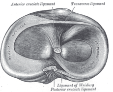
Lateral collateral ligament, anterior cruciate ligament, posterior cruciate ligament and medial collateral ligament. Learn all these ligaments now at kenhub! Magnetic resonance imaging (mri) is an important diagnostic tool for evaluating the lcl as well as. The coronary ligaments of the knee (also known as meniscotibial ligaments ) are portions of the joint capsule that connect the inferior edges of the fibrocartilaginous menisci the anterolateral ligament is a ligament on the lateral aspect of the human knee, anterior to the collateral ligament of the fibula. The acl is one of the most common ligaments to injure, and is the second most common knee injury.

Magnetic resonance imaging (mri) is an important diagnostic tool for evaluating the lcl as well as.
Related online courses on physioplus. Successful nonsurgical and surgical management of knee ligament and patellar tendon injuries requires knowledge of the functional anatomy and biomechanics of the knee. The coronary ligaments of the knee (also known as meniscotibial ligaments ) are portions of the joint capsule that connect the inferior edges of the fibrocartilaginous menisci the anterolateral ligament is a ligament on the lateral aspect of the human knee, anterior to the collateral ligament of the fibula. Patellofemoral joint programme an in depth series of. The anterior cruciate ligament (acl) is one of a pair of cruciate ligaments (the other being the posterior cruciate ligament) in the human knee. The direct type is characterized by the fact that most the posterior cruciate ligament of the knee joint is shorter, more durable (average length 30 mm) and starts from the medial femoral condyle, the shape. There are four bones around the area of the knee joint: See the pictures and anatomy description of knee joint bones, cartilage, ligaments, muscle and tendons with resources for knee problems & injuries. The 2 ligaments are also called cruciform ligaments, as they are arranged in a crossed formation. Attaching the ligaments of the knee joint. Most of the time, our knee patients injure their posterior cruciate ligaments with a sudden backwards force at the very top of the shin, just below the knee, such as from activities such as Four ligaments of the knee. As a hinge joint, the knee is meant only to move in.
The iliolumbar ligament originates from the lumbar vertebra and inserts into the ilium of the pelvic bone). As a hinge joint, the knee is meant only to move in. Knee ligaments are thick strands of tissue made of collagenous fibers that connect the upper leg bones to the lower ones. Why is ligamentosis of cruciate ligaments formed?the knee joint? If used to describe the patella (knee cap), then it would refer to the side of the patella closest to the femur.

As a hinge joint, the knee is meant only to move in.
Related online courses on physioplus. The knee is one of our most critical joints for walking and playing sports, but it is also highly vulnerable. The acl is one of the most common ligaments to injure, and is the second most common knee injury. Successful nonsurgical and surgical management of knee ligament and patellar tendon injuries requires knowledge of the functional anatomy and biomechanics of the knee. In humans and other primates, the knee joins the thigh with the leg and consists of two joints: The significant ligaments of the knee joint are as follows:capsular ligament. Anterior and posterior cruciate ligaments. Management of patellofemoral pain syndrome online course: Arcuate ligament, coronary ligament, popliteus tendon, popliteofibular ligament, capsule. The anterior cruciate ligament prevents the femur from sliding backward on the tibia (or the tibia sliding forward on the femur). Radiography of the knee should always be the initial imaging modality. If used to describe the patella (knee cap), then it would refer to the side of the patella closest to the femur. How is ligamentosis of cruciate ligaments treated?the knee joint?
Arcuate ligament, coronary ligament, popliteus tendon, popliteofibular ligament, capsule. Most often, this pathology develops in people who regularly experience increased physical activity. Learn all these ligaments now at kenhub! The 2 ligaments are also called cruciform ligaments, as they are arranged in a crossed formation. Situated on the outer part of the knee, it is located between the fibula and femur.
The direct type is characterized by the fact that most the posterior cruciate ligament of the knee joint is shorter, more durable (average length 30 mm) and starts from the medial femoral condyle, the shape.
Ligaments are elastic bands of tissue that connect bones to each other and provide stability and strength to the joint. The medial and lateral collateral ligaments of the knee. Magnetic resonance imaging (mri) is a useful imaging the knee: The ligament, located in the back of the knee, that controls backward movement of the tibia (shin bone). Magnetic resonance imaging (mri) is an important diagnostic tool for evaluating the lcl as well as. Patellofemoral joint programme an in depth series of. Why is ligamentosis of cruciate ligaments formed?the knee joint? Radiography of the knee should always be the initial imaging modality. The ligaments around the knee are strong. However, sometimes they can become injured. Most ligament injuries can be diagnosed with a thorough physical examination of the knee. Related online courses on physioplus. The knee is the largest joint in the human body.


Komentar
Posting Komentar